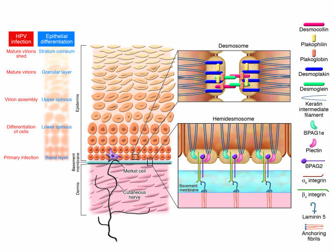
Figure 1. Schematic illustration of several key elements of the skin.
As indicated, the skin is divided into 2 main compartments, epidermal (tan) and dermal (pink), separated by a basement membrane (blue). The epidermis serves as the protective barrier, due to the differentiation of proliferative epithelial cells in the basal layer to the terminally differentiated cells in the stratum corneum. The sites and development of HPV infection are indicated at far left: the primary infection occurs in the basal layer, and mature virion sheds in the stratum corneum. Merkel cells are located within the basal layer of the epidermis (purple) and are associated with sensory nerve endings (brown). Two types of cell junctions critical for the integrity of the skin are highlighted here. Desmosomes (upper right inset) form cell-cell junctions, in which cadherins (pink and green), such as desmogleins 1 and 3, are the extracellular bridges and the autoantigens in different forms of pemphigus. Hemidesmosomes (lower right inset) tether the cells to the basement membrane and are composed of a number of proteins, 2 of which — BP230 and BP180 (also known as type XVII collagen) — are autoantigens in BP. Disruption of these interactions results in loss of adhesion of the cells to one another or to the underlying basement membrane.

