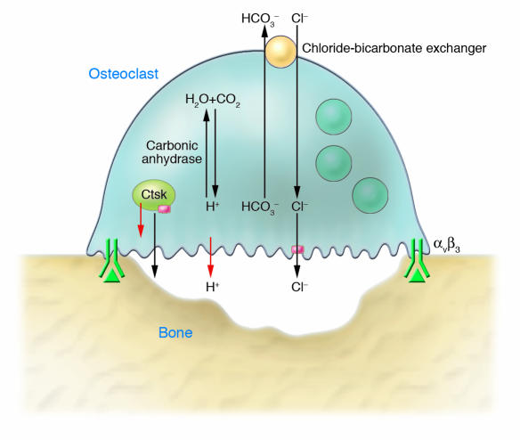
Figure 3. Mechanism of osteoclastic bone resorption.
The osteoclast adheres to bone via binding of RGD-containing proteins (green triangle) to the integrin αvβ3, initiating signals that lead to insertion into the plasma membrane of lysosomal vesicles that contain cathepsin K (Ctsk). Consequently, the cells generate a ruffled border above the resorption lacuna, into which is secreted hydrochloric acid and acidic proteases such as cathepsin K. The acid is generated by the combined actions of a vacuolar H+ ATPase (red arrow), its coupled Cl– channel (pink box), and a basolateral chloride–bicarbonate exchanger. Carbonic anhydrase converts CO2 and H2O into H+ and HCO3–. Solubilized mineral components are released when the cell migrates; organic degradation products are partially released similarly and partially transcytosed to the basolateral surface for release.

