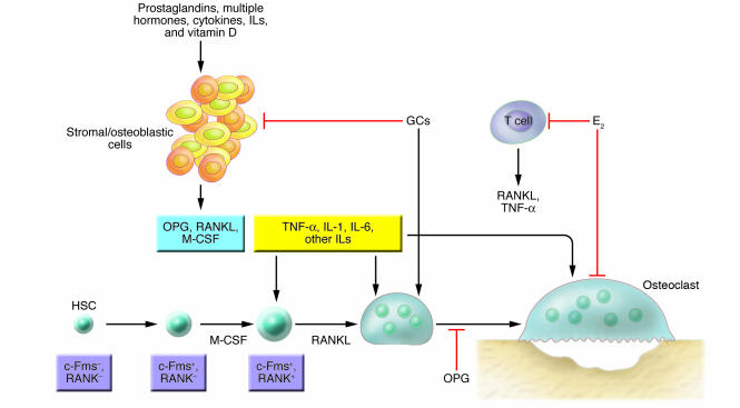
Figure 4. Role of cytokines, hormones, steroids, and prostaglandins in osteoclast formation.
Under the influence of other cytokines (not shown), an HSC commits to the myeloid lineage, expresses the M-CSF receptor colony-stimulating factor receptor 1 (c-Fms) and then, driven by M-CSF/c-Fms signaling and the RANKL receptor RANK, differentiates into an osteoclast. Mesenchymal cells in the marrow respond to a range of hormonal and cytokine stimuli, secreting a mixture of pro- and antiosteoclastogenic proteins, the latter primarily being OPG. Glucocorticoids (GCs) suppress bone resorption indirectly (by inducing death of osteoblasts) but possibly also target osteoclasts and/or their precursors. Estrogen (E2) inhibits secretion of RANKL and TNF-α by T cells via a complex mechanism (not shown); the sex steroid also inhibits osteoclast function.

