
Figure 2.
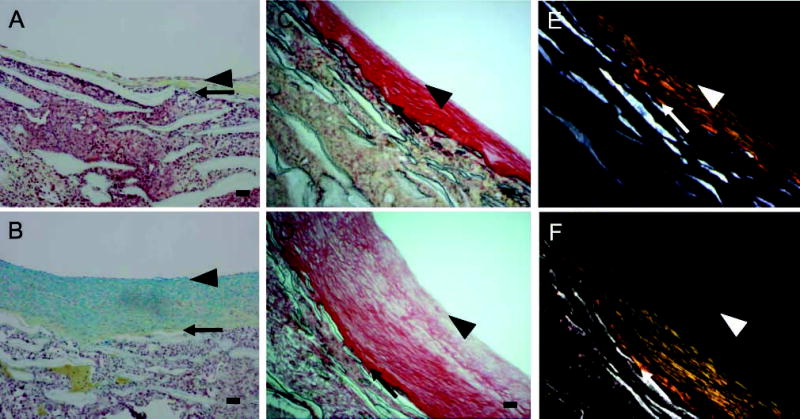
Movat’s pentachrome (A,B) and Sirius Red (C–F) staining of aorto-iliac-PTFE grafts healed for 12 weeks under high flow (A,C,E) or for 8 weeks under high flow and 4 weeks under normal flow (B,D,F). Sirius Red staining was visualized under normal (C,D) or polarizing (E,F) light. Arrows indicate the intima/PTFE boundary, while the arrowheads indicate the subendothelial layer. The Movat’s pentachrome procedure stains proteoglycans blue, collagen yellow, elastin black, and cells red. Bars = 0.1 mm.