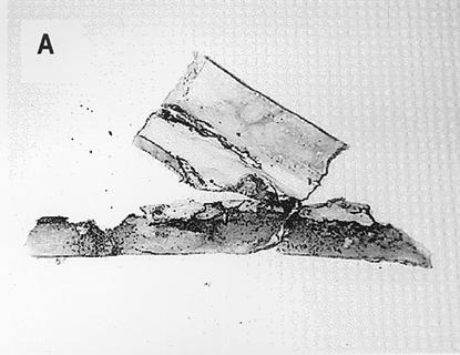
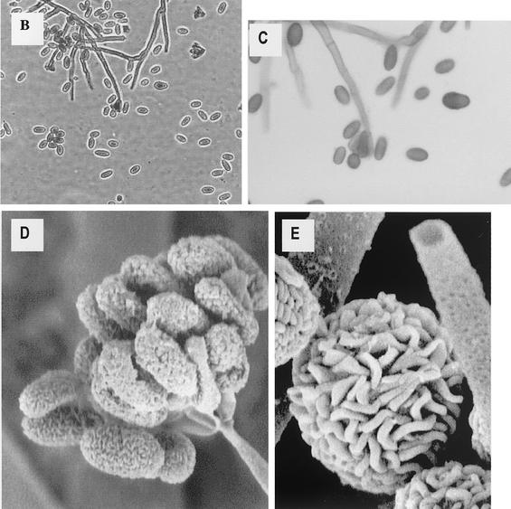
FIG. 1.
Morphology of S. chartarum. (A) Representative section of damaged wallboard. Note the areas of black discoloration. (B) Pure culture of S. chartarum, initially obtained from contaminated wallboard (40× objective). (C) Higher-power view of the same culture as in panel B (100× oil objective). (D) Scanning electron micrograph of conidia at the tip of a conidiaphore. (E) Scanning electron micrograph of mature conidia. Slime has been removed by scanning electron microscope processing. Panels D and E are reprinted from Nelson, http://www.apsnet.org/online/feature/stachybotrys/ with permission.


