
FIG. 4.
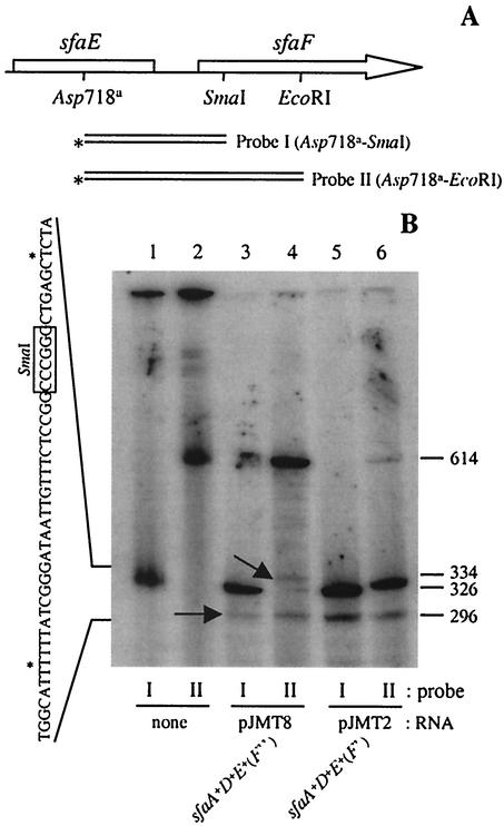
The 3′ end of the sfaADE transcript mapped in the coding sequence of sfaF. (A) Genetic map of the DNA fragments used as probes for S1 nuclease digestion. The asterisks indicate the 3′end that was radiolabeled. (B) S1 nuclease digestion of total RNA from strains HB101/pJMT8 (lanes 3 and 4) and HB101/pJMT2 (lanes 5 and 6) by using probe I (lanes 1, 3, and 5) and probe II (lanes 2, 4, and 6). The arrows indicate the positions of two different kinds of products obtained. On the left side the exact positions of the two 3′ ends of the sfaADE transcript are indicated (asterisks) in the sequence. The boxed nucleotides indicate the position of the SmaI restriction site. The exact size (number of nucleotides, shown on the right) of the S1 products was determined by using sequencing reaction mixtures as molecular mass markers (data not shown).