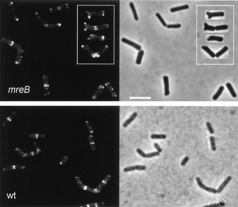
FIG. 3.
Localization of DivIVA-GFP under MreB-depletion conditions. The left upper panel (mreB) shows fluorescence images of B. subtilis strain 3292 cells grown in Spizizen's minimal medium without xylose. The inset shows some examples of cells with deformed poles. Scale bar in right half of panel, 5 μm. The left lower panel (wt) shows fluorescence images of wild-type B. subtilis (strain 1803) grown under the same conditions. Corresponding phase-contrast images are shown in the right halves of the panels.

