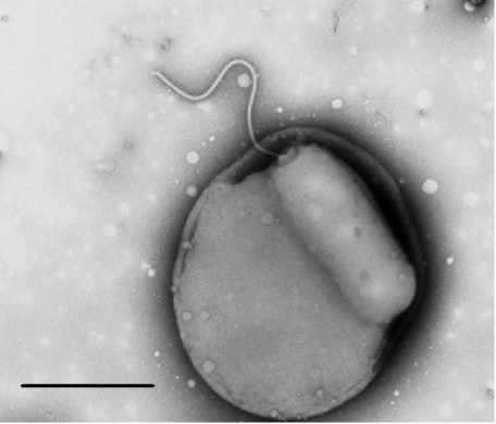
Fig. 1.
Electron micrograph of B. bacteriovorus wild-type strain 109J inside a bdelloplast of a small E. coli DFB225 flagellar minus cell upon which it is preying, 20 min after predators were added to prey. The Bdellovibrio has modified the cell wall of the prey, which has rounded up, and it is attached to the cytoplasmic membrane of the prey, consuming its cytoplasm. The flagellum of the Bdellovibrio is still clearly visible protruding from the bdelloplast, 1% PTA stain. Bar = 1 µm.

