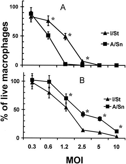
FIG. 3.
Interstrain differences in viability of I/St and A/Sn peritoneal and lung macrophages following in vitro infection with M. tuberculosis H37Rv. Peritoneal (A) and lung (B) macrophages were isolated, purified, plated, and infected (see Materials and Methods). The number of cells that remained adherent and retained the appearance of uninfected control macrophages from the same source, as well as the total number of cells, were determined using an inverted microscope equipped with a 10-mm2 grid in the eyepiece. Three random areas per well and three wells per condition were counted at ×100 magnification with estimation of the mean. The results obtained in four independent experiments at 72 h of culture are displayed as an aggregate mean of the percentage of live cells ± SD. Interstrain differences were statistically significant (P < 0.01 to 0.05; Mann-Whitney U test) at MOIs of 0.6 to 2.5 for peritoneal macrophages and 1.2 to 10.0 for lung macrophages (marked with asterisks). Similar results, although with statistically significant differences shifted to higher MOI values (2.5 to 10.0), as expected, were obtained in 48-h cultures.

