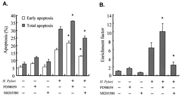
FIG. 3.
Augmentation of H. pylori-induced apoptosis by inhibition of ERK1/2 pathway. (A) H. pylori strain 99 (cagA+, cytotoxin positive) and a subconfluent monolayer of AGS cells were cocultured for 16 h with or without pretreatment of PD98059 (20 μM) or SB203580 (10 μM). Data represent early and total apoptotic cell proportions measured by flow cytometric analysis after staining with FITC-conjugated annexin V and propidium iodide. (B) Apoptosis of AGS cells was assessed by a cell death detection ELISA. Values refer to DNA fragmentation as measured by the enrichment factor. Values are expressed as means ± standard deviations of a triplicate experiment and are representative of three separate experiments. *, P < 0.05 (compared with results for H. pylori infection alone).

