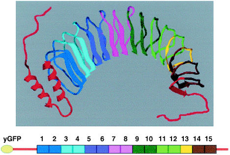
FIG. 1.
Structure of YopM and cartoon of yEGFP-YopM. (Top) Ribbon model of YopM based on PDB structure 1JL5 (12) modified by adding free-form lines to indicate residues at the beginning of the leader domain (red, at left) and at the end of the tail domain (red, at right) which were not resolved in the crystal structure. Coloring has been added to provide a visual aid for locating regions deleted in the various yEGFP-YopM molecules tested in this study (Table 1). (Bottom) Cartoon of yEGFP-YopM with numbered LRRs and the same coloring as in the ribbon model. The figure was generated using Swiss PBD Viewer and rendered with PovRay before final construction in Microsoft Powerpoint. It was printed from Adobe PhotoShop 6.0.

