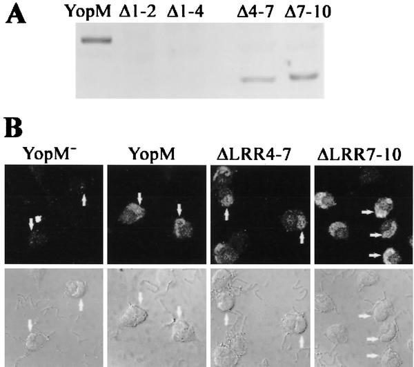
FIG. 5.
YopM ΔLRR4-7 and YopM ΔLRR7-10 also localize to the nucleus in HeLa cells after delivery by Y. pestis infection. HeLa cells were infected for 4 h with YopM− Y. pestis KIM8-3233 or Y. pestis KIM8-3233 expressing YopM, YopM ΔLRR1-2, YopM ΔLRR1-4, YopM ΔLRR4-7, or YopM ΔLRR7-10. After a brief trypsin treatment, the infected cells were lysed with water to obtain the soluble cellular fraction (cytosol). YopM proteins that had been delivered to the HeLa cytosol by the surface-adherent yersiniae were visualized by immunoblot analysis and probed with a rabbit polyclonal antibody raised against the whole YopM, which recognizes all of the YopM proteins being tested. Lanes: YopM, wild-type YopM; Δ1-2, YopM ΔLRR1-2; Δ1-4, YopM ΔLRR1-4; Δ4-7, YopM ΔLRR4-7; Δ7-10, YopM ΔLRR7-10. (B) The distribution of YopM protein in the infected HeLa cells was determined by indirect immunofluorescence. The primary antibody was the rabbit polyclonal used in panel A; the secondary antibody was conjugated to the Oregon Green fluorochrome. The upper panels show Oregon Green fluorescence from an optical slice obtained by laser scanning confocal microscopy; the lower panels show the differential interference contrast image of the same cells. From left to right, the panels illustrate two, two, five, and six infected HeLa cells. The arrows on some of the cells point to the kidney bean-shaped nucleus that lies to one side of each cell.

