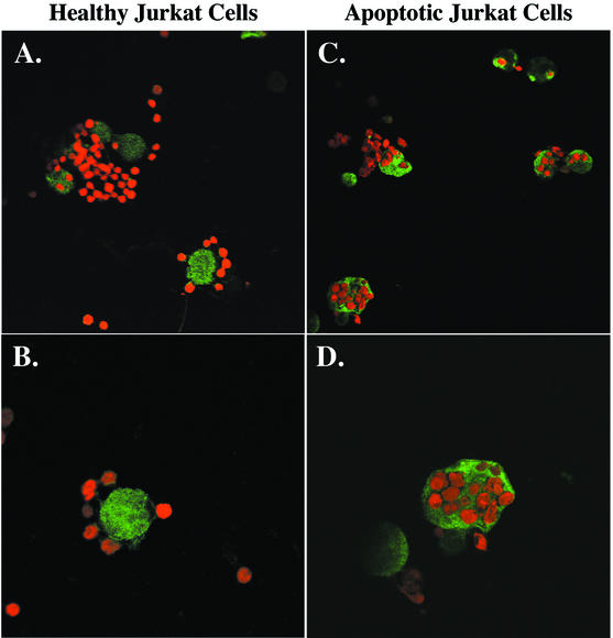
FIG. 2.
Confocal microscopy of the interaction of healthy and previously killed Jurkat cells with NH4Cl-treated E. histolytica trophozoites. TAMRA-labeled healthy or apoptotic Jurkat cells (red) were centrifuged onto NH4Cl-pretreated E. histolytica trophozoites (E. histolytica/Jurkat cell ratio, 1:4). Following 10 min of incubation (37°C), unbound cells were washed away, and remaining cells were fixed and stained for E. histolytica Gal/GalNAc lectin with rabbit antilectin polyclonal antibodies and FITC-conjugated anti-rabbit mouse antibody (green). Original magnifications, A and C, ×400; B and D, ×800.

