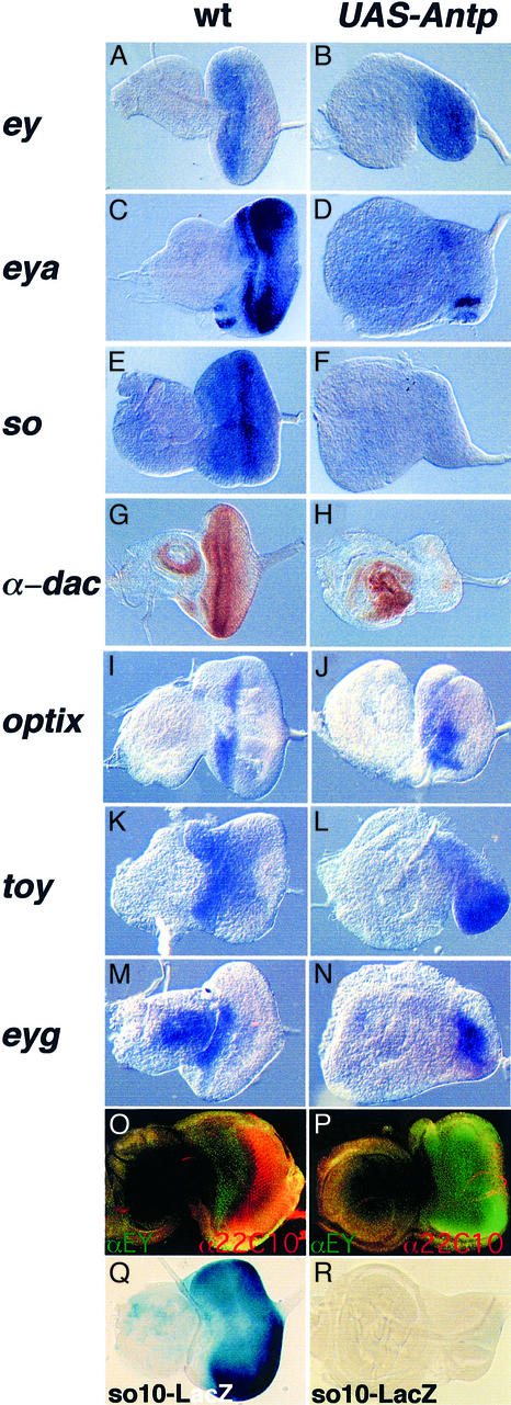
Fig. 2. ANTP represses the ey regulatory pathway and blocks photoreceptor differentiation despite the presence of EY. In situ hybridization or immunostaining (G, H, O and P) experiments were performed on eye antenna third instar imaginal discs to study gene expression following Antp expression. (A, C, E, G, I, K, M, O and Q) Wild-type discs; (B, D, F, H, J, L, N, P and R) targeted expression of Antp with dppblink-Gal4. The magnification is 2-fold higher for the ANTP-expressing disc as compared with the wild type. (O and P) Immunostaining experiment using an αEY antibody (in green) and the α22C10 neuronal marker (in red). Note, in wild type (O) the expression of EY is restricted anterior to the furrow. (Q and R) Analysis of the EY responsive element so10 enhancer expression in wild-type and Antp-expressing discs. β-galactosidase staining was performed in parallel in wild-type (Q) as well as in Antp-expressing discs (R).

An official website of the United States government
Here's how you know
Official websites use .gov
A
.gov website belongs to an official
government organization in the United States.
Secure .gov websites use HTTPS
A lock (
) or https:// means you've safely
connected to the .gov website. Share sensitive
information only on official, secure websites.
