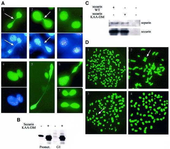
Fig. 8. Expression of indestructible securin interferes with chromatid separation. (A) NIH 3T3 cells were transfected with vectors expressing non-degradable (panels 1, 2, 4, 5 and 6) and wild-type (panel 3) securin together with a GFP–histone H2A expression vector (green panels). Cells were stained further in vivo with Hoechst 33258 (blue panels). About 1/3 of the cells expressing the mutant securin failed to separate all their chromatids prior to cytokinesis and remained connected by a thin chromatin string (arrow), or even failed to complete nuclear division altogether (panel 4). The same phenotype was observed in some HeLa cells expressing non-degradable securin (panel 7). (B) Cells transfected with an empty vector or with non-degradable securin were arrested in prometaphase and either harvested immediately or released first into G1 for 3 h. Cell extracts subsequently were immunoblotted with securin antibodies. (C) Cells were transfected with an expression vector for myc-tagged wild-type and non-degradable securin. Extracts prepared from transfected and untransfected cells were immunoprecipitated with myc tag (9E10) antibodies and immunoblotted with separin and securin antibodies. (D) Cells were co-transfected with expression vectors for securin (wild-type or non-degradable), non-degradable cyclin B1 and GFP–H2A. Cells were arrested in mitosis and were harvested by shake-off. Chromosome spreads were prepared as described in Materials and methods. Cells transfected with wild-type, and the majority of those transfected with non-degradable securin, completely dissociated chromatids (panel 1). However, many of those transfected with non-degradable securin still had a few (panel 2; arrow shows a separated chromosome), most (panel 3; arrow shows an unseparated chromosome) or all (panel 4) chromatids paired.

An official website of the United States government
Here's how you know
Official websites use .gov
A
.gov website belongs to an official
government organization in the United States.
Secure .gov websites use HTTPS
A lock (
) or https:// means you've safely
connected to the .gov website. Share sensitive
information only on official, secure websites.
