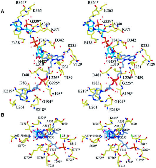
Fig. 5. Ligand-binding sites. (A) Stereoview of the binding site for FAD and NADPH. The carbon atoms of the cofactor FAD are shown in gray, those of the cosubstrate NADPH in cyan. Side chains that are conserved between pig liver DPD and B.taurus adrenodoxin reductase are marked with an asterisk. (B) Stereoview of the substrate-binding site shows 5FU bound adjacent to the cofactor FMN in the C671A mutant. A 2|Fo| – |Fc| map is contoured at 1.2σ for 5FU. Side chains that are conserved between pig liver DPD and L.lactis DHOD(A) are marked with an asterisk.

An official website of the United States government
Here's how you know
Official websites use .gov
A
.gov website belongs to an official
government organization in the United States.
Secure .gov websites use HTTPS
A lock (
) or https:// means you've safely
connected to the .gov website. Share sensitive
information only on official, secure websites.
