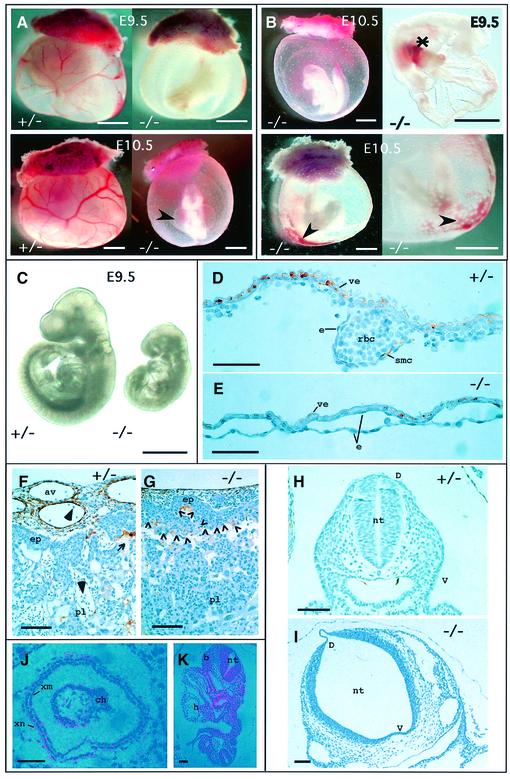
Fig. 2. TβRI–/– embryos exhibit severe defects in vascular development. (A) Gross morphology of whole-mount yolk sacs in mutant embryos compared with heterozygous littermates. Note the enlarged pericardial sac (arrowhead). (B) Defects in TβRI–/– embryos. Some embryos have not turned (upper left). Asterisk, accumulated blood in vitilline vessels connecting the yolk sac and the embryo proper. Arrowheads, leakage of red blood cells and dilated vascular structures. (C) By E9.5 mutant embryos are severely growth retarded. (D–G) Immunohistochemical staining for smooth muscle cell actin in transverse sections of mutant embryos compared with heterozygous littermates. (D) Section through the yolk sac of a heterozygote at E9.5. Endothelial cells are either in close apposition with the visceral endoderm or contain red blood cells within the developing vessel. Smooth muscle cells, stained brown, begin to differentiate from mesenchyme and contribute to the structure of the vessel wall. (E) Section through the yolk sac of a mutant at E9.5. The vessels are lined with endothelial cells but are dilated, and have very few red blood cells and no smooth muscle cells. (F) Section through chorioallantoic region of the placenta of a heterozygote at E9.5 showing smooth muscle cells surrounding the dilated allantoic blood vessels derived from extra-embryonic tissue and their invasion of the labyrinthine part of the placenta (maternal part). This allows intermingling of the fetal (large arrowheads) and maternal (small arrow) red blood cells. (G) Allantoic blood vessels in the placenta of a mutant at E9.5 are much smaller, there are fewer smooth muscle cells and there is no evidence for their invasion of the maternal labyrinthine layer through the ectoplacental plate. The limit of ectoplacental plate is indicated by the row of open arrowheads in (G). (H and I) Histological sections of the neural tube at the level of the heart in a heterozygote (H) and homozygote (I) at E8.5. (J) Expression of TβRI at E7.5 in the extra-embryonic part of the conceptus. (K) Expression of TβRI in the rostral neural tube at the level of the heart at E8.5. Abbreviations: av, allantoic blood vessel; b, branchial arch; ch, chorion; D, dorsal; e, endothelial cell; ep, ectoplacental plate; h, heart; nt, neural tube; pl, placenta; rbc, red blood cell; smc, smooth muscle cells; V, ventral; ve, visceral endoderm; xm, extra-embryonic mesoderm; xn, extra-embryonic endoderm. Scale bars, 1 mm (A–C), 50 µm (D–E), 100 µm (F–K).

An official website of the United States government
Here's how you know
Official websites use .gov
A
.gov website belongs to an official
government organization in the United States.
Secure .gov websites use HTTPS
A lock (
) or https:// means you've safely
connected to the .gov website. Share sensitive
information only on official, secure websites.
