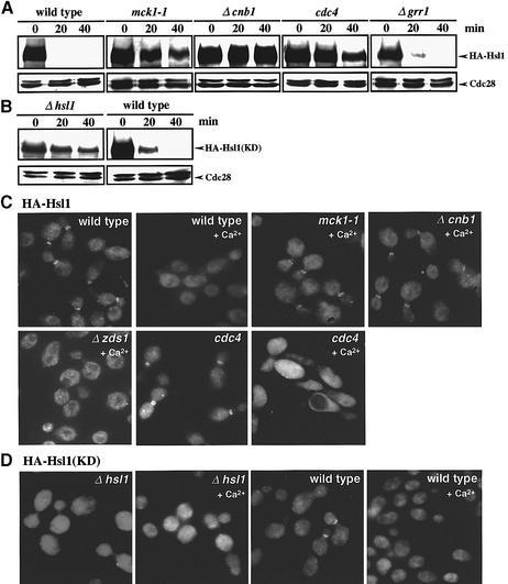
Fig. 6. Ca2+ induces SCFCdc4-mediated Hsl1 destabilization and Hsl1 delocalization from the bud neck in a manner dependent on both calcineurin and Mck1. (A) Comparison of Hsl1 abundance in various strains. Wild type (YMM53), mck1-1 (YMM71), Δcnb1 (YMM72), Δzds1 (YMM54), cdc4 (YMM73) or Δgrr1 (YMM76), each bearing a gene encoding HA-Hsl1, was grown in liquid YPD at 28°C until early log phase, and then shifted to 37°C for 2 h to inactivate Cdc4. CaCl2 was added to the cell cultures to a final concentration of 100 mM, samples were taken at intervals of 20 min and western blot analysis was performed. (B) Comparison of the Ca2+-induced destabilization of a kinase-negative Hsl1, HA-Hsl1(KD), in strains Δhsl1 (YMM60) or wild-type HSL1 (YMM66), each bearing a gene encoding HA-Hsl1(KD). Experimental conditions were as described in (A). (C) HA-Hsl1 localization in various strains. The cells were grown in liquid YPD at 28°C until early log phase, and then shifted to 37°C for 2 h to inactivate cdc4. CaCl2 was added to the cell cultures to a final concentration of 100 mM, and samples were then taken after 20 min of incubation. Indirect immunofluorescence microscopy was performed as described in Materials and methods. (D) Comparison of the localization of HA-Hsl1(KD) in Δhsl1 (YMM60) or HSL1 (YMM66) cells. CaCl2 was added to the cell cultures to a final concentration of 100 mM, and samples were then taken after 20 min of incubation. Experimental conditions were the same as in (C).

An official website of the United States government
Here's how you know
Official websites use .gov
A
.gov website belongs to an official
government organization in the United States.
Secure .gov websites use HTTPS
A lock (
) or https:// means you've safely
connected to the .gov website. Share sensitive
information only on official, secure websites.
