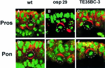
Fig. 2. Localization of cell fate determinants is defective in deficiency lines Df(2L)osp29 and Df(2L)TE35BC-3. Confocal images of dividing NBs double-labelled with anti-Pros (red, A–C) or anti-Pon (red, D–F) and DNA staining to indicate the condensed chromosomes (green). Note that in wild-type embryos (A and D), Pros and Pon form basal crescents in dividing neuroblasts, while in the deficiency lines, although the crescents are formed, they fail to move to the basal cortex (B, C, E and F). Apical is up. NBs are outlined with white dots.

An official website of the United States government
Here's how you know
Official websites use .gov
A
.gov website belongs to an official
government organization in the United States.
Secure .gov websites use HTTPS
A lock (
) or https:// means you've safely
connected to the .gov website. Share sensitive
information only on official, secure websites.
