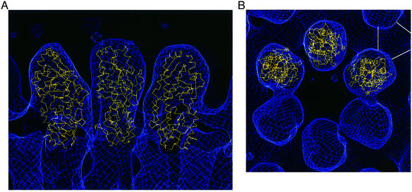
Fig. 6. Fit of the structure in the low-resolution map of the virion from electron cryomicroscopy of a type 1 Lang (T1L) virion (S.B.Walter and T.S.Baker, personal communication). (A) A cross-section of a ring of six σ3 proteins on the surface of the virion, with a view tangential to the curvature of the virion, and the center of the virion below the figure. (B) A view looking down on the surface of a virion. The virion map is shown in blue, and σ3 models are displayed as α-carbon backbone traces in yellow. The orientation of the central model in (A) is the same as that in Figure 1B, with the small lobe on the bottom. The white lines in the upper right hand corner of (B) indicate a trimer of the underlying protein, µ1.

An official website of the United States government
Here's how you know
Official websites use .gov
A
.gov website belongs to an official
government organization in the United States.
Secure .gov websites use HTTPS
A lock (
) or https:// means you've safely
connected to the .gov website. Share sensitive
information only on official, secure websites.
