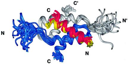
Fig. 3. Similarity of the AKAP structure and binding region on RIIα(1–44) in the Ht31 and AKAP79 complexes. The superposition in RIIα(1–44), with the protomers of RIIα D/D shown in gray and blue, reveals the similarity in the backbone fold of the two complexes. The first 17 residues of the RII binding region in both Ht31(493–515) (red) and AKAP79(392–413) (yellow) are indicated, and bind to identical regions with similar helical structures to RIIα(1–44).

An official website of the United States government
Here's how you know
Official websites use .gov
A
.gov website belongs to an official
government organization in the United States.
Secure .gov websites use HTTPS
A lock (
) or https:// means you've safely
connected to the .gov website. Share sensitive
information only on official, secure websites.
