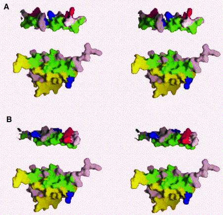
Fig. 6. Stereoview representation of the AKAP binding surface of RIIα using Insight software (MSI, San Diego, CA). RIIα(1–44) protomers are colored yellow and pink, respectively. All hydrophobic residues in RIIa, Ht31 and AKAP79 are colored green. Acidic and basic residues are red and blue, respectively. (A) Ht31 lies above RIIα, with the hydrophobic face that contacts RIIα facing the hydrophobic face of RIIα. Additional residues in Ht31 are colored pink. (B) AKAP79 is shown in this figure with the hydrophobic face in similar orientation to RIIα to that of Ht31 in (A). Additional residues in Ht31 are colored pink.

An official website of the United States government
Here's how you know
Official websites use .gov
A
.gov website belongs to an official
government organization in the United States.
Secure .gov websites use HTTPS
A lock (
) or https:// means you've safely
connected to the .gov website. Share sensitive
information only on official, secure websites.
