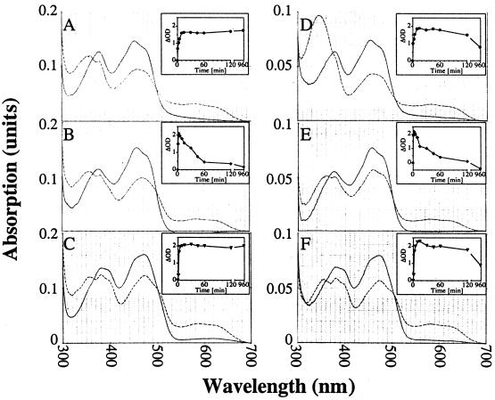
Figure 1.
Visible absorption spectra of cytochrome P450 reductase and mutants. Semiquinone spectra of sCPR (A, D), sW676H (B, E) and sW676A (C, F) were obtained after the addition of a 5-fold molar excess of NADPH (A–C) or NADH (D–F) under aerobic conditions, and recorded after equilibration has been reached. The reoxidation of the semiquinone (Insets) was followed at 585 nm for 16 h (solid lines: oxidized spectra; broken lines: reduced spectra).

