Figure 1.
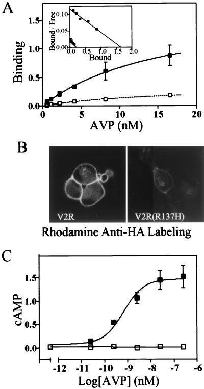
Expression and adenylyl cyclase stimulation of V2R and V2R(R137H) in HEK-293 cells. Cells transiently transfected with cDNA for V2R (■) or V2R(R137H) (□) were exposed to varying concentrations of [3H]AVP. (A) Scatchard analysis (Inset) indicates the receptors have similar affinity for AVP [V2R, 16 ± 6 nM; V2R(R137H), 15 ± 3 nM]. V2R expression varied between 2.5 and 5 pmol/mg of cell protein, with the plasma membrane expression of the V2R(R137H) being approximately 1/12 of this (x intercept of Scatchard). The data are representative of three experiments, with each point measured in duplicate. (B) Fluorescence images of live unpermeabilized cells, labeled with rhodamine-tagged mouse-monoclonal anti-HA antibodies, expressing the V2R (Left) or the V2R(R137H) (Right). (C). cAMP measured in whole cells stimulated for 15 min with concentrations of AVP between 0 and 250 nM. cAMP accumulation is expressed as total counts of [3H]cAMP/total uptake of [3H]adenine per well of cells. Results are the mean ± SD of three experiments.

