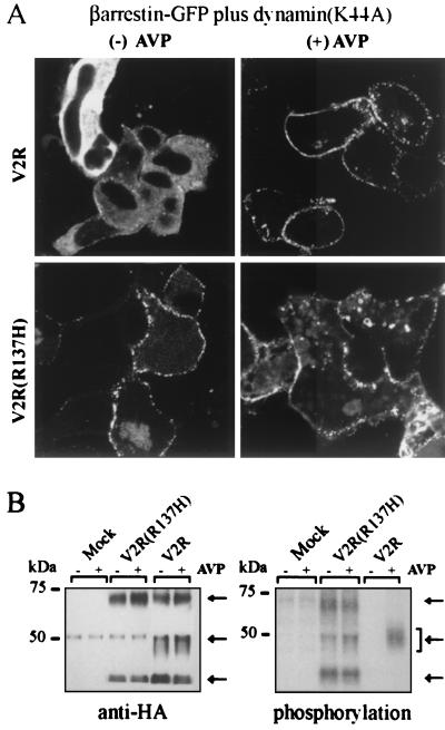
Figure 4.
βarrestin2 association with and phosphorylation of V2R and V2R(R137H) in HEK-293 cells. (A) Dynamin(K44A) was expressed with βarrestin2-GFP and either V2R or V2R(R137H). Exposure of V2R to AVP (100 nM) (Upper Right) resulted in appreciable βarrestin2-GFP translocation that remains visible at 30 min as a punctate distribution at the cell membrane rather than as a vesicular distribution inside the cell. In the absence of agonist, the cells containing dynamin(K44A) and V2R(R137H) (Lower Left) also show βarrestin2-GFP fluorescence distributed in punctate areas at the plasma membrane. A similar pattern was apparent even in the presence of agonist (Lower Right). (B) Left shows receptors that were immunoprecipitated with a mouse anti-HA antibody and blotted with a rabbit anti-HA antibody. The faint 50-kDa band present in all six lanes is cross-reactive mouse Ig heavy chain. Right shows depicts receptors that were assayed for phosphorylation as described in Experimental Procedures. Equal amounts of receptor (40 fmol) were loaded into each lane. The arrows mark the positions of the receptor species migrating at approximately 70, 50, and 40 kDa as revealed by anti-HA antibody. Results are representative of three experiments.

