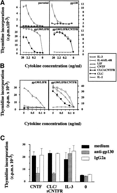
Fig. 5. Proliferative response of transfected Ba/F3 cell lines to the CLC–sCNTFR complex. (A) Effect of CLC–sCNTFR on parental Ba/F3 cells and cells transfected with gp130, gp130–LIFR or gp130–LIFR–CNTFR. Cells were cultured in triplicate with 3-fold dilutions of appropriate positive controls, purified CLC–sCNTFR (filled squares) or IL-2 (open circles) used as an irrelevant cytokine. After a 72 h culture period, a [3H]Tdr pulse was performed and the amount of incorporated radioactivity was determined using a β-counter. Vertical bars indicate the SEM. (B) Effect of purified CLC from Cos-7 transfected cell lysates on Ba/F3 cells transfected with gp130–LIFR and gp130–LIFR–CNTFR. Cells were cultured in triplicate with 3-fold dilutions of the indicated cytokines. (C) The AN-HH1 anti-gp130 mAb prevents the proliferative response of the Ba/F3 gp130/LIFR/CNTFR cell line to the CLC–sCNTFR complex. Transfected Ba/F3 cells were incubated in triplicate in culture medium (marked as 0) containing 1 ng/ml CNTF, CLC–sCNTFR or IL-3. The AN-HH1 antibody (hatched bars) or a control IgG2a antibody (open bars) was added at a final concentration of 30 µg/ml. After a 72 h culture period, [3H]Tdr was added for 4 h, and the incorporated radioactivity determined.

An official website of the United States government
Here's how you know
Official websites use .gov
A
.gov website belongs to an official
government organization in the United States.
Secure .gov websites use HTTPS
A lock (
) or https:// means you've safely
connected to the .gov website. Share sensitive
information only on official, secure websites.
