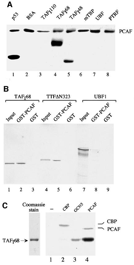
Fig. 3. PCAF acetylates TAFI68 in vitro. (A) Acetylation of recombinant proteins. A 2 µg aliquot of the proteins indicated was incubated with 500 ng of FLAG-PCAF, 1 µCi of [3H]acetyl-CoA and 0.4 µM TSA in a total volume of 30 µl of buffer AM-100 for 30 min at 30°C. Proteins were separated by 10% SDS–PAGE, and acetylated proteins were visualized by fluorography. (B) TAFI68 interacts with PCAF. GST–PCAF or GST were bound to glutathione–agarose beads and incubated with 35S-labeled TAFI68 (lanes 2 and 3), TTFΔN323 (lanes 5 and 6) or UBF (lanes 8 and 9). Bound proteins were analyzed by 8% SDS–PAGE and autoradiography. Ten percent of the 35S-labeled input proteins are shown in lanes 1, 4 and 7. (C) Acetylation of TAFI68 with CBP, GCN5 and PCAF. A 500 ng aliquot of TAFI68 was incubated for 30 min at 30°C with 1 µCi of [3H]acetyl-CoA, 0.4 µM TSA and comparable units of HAT activity of CBP (lane 2), GCN5 (lane 3) or PCAF (lane 4). After gel electrophoresis, acetylated TAFI68 was visualized by fluorography. A Coomassie Blue stain of 500 ng of TAFI68 is shown on the left.

An official website of the United States government
Here's how you know
Official websites use .gov
A
.gov website belongs to an official
government organization in the United States.
Secure .gov websites use HTTPS
A lock (
) or https:// means you've safely
connected to the .gov website. Share sensitive
information only on official, secure websites.
