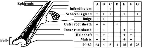
Fig. 2. Distribution of lacZ-positive cells in various compartments of labeled pilosebaceous units. Serial sections of skin of transduced mice following five cycles of induced hair growth, at 37 weeks post-transduction, were analyzed by X-gal staining and the distribution of lacZ-positive clusters in the various compartments of 82 pilosebaceous units was noted. ‘+’ indicates partial or uniform labeling of cells in that compartment. The number of labeled pilosebaceous units in each category is shown in the bottom row.

An official website of the United States government
Here's how you know
Official websites use .gov
A
.gov website belongs to an official
government organization in the United States.
Secure .gov websites use HTTPS
A lock (
) or https:// means you've safely
connected to the .gov website. Share sensitive
information only on official, secure websites.
