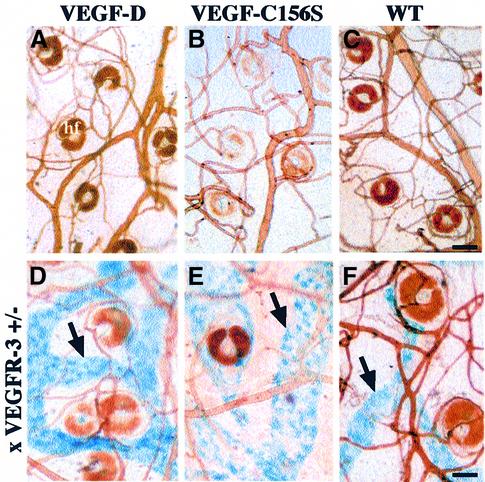
Fig. 4. Whole-mount analysis of skin blood and lymphatic vasculature. Blood vessels were visualized by injecting the mice intravenously with biotin-labelled L.esculentum lectin followed by vascular perfusion (A–F). Lymphatic vessels were stained blue (arrows) in the skin of K14-VEGF-D and K14-VEGF-C156S mice crossed with heterozygous mutant VEGFR-3+/LacZ mice (D–F). Scale bar in (C) for (A–C), 75 µm; in (F) for (D–F), 65 µm.

An official website of the United States government
Here's how you know
Official websites use .gov
A
.gov website belongs to an official
government organization in the United States.
Secure .gov websites use HTTPS
A lock (
) or https:// means you've safely
connected to the .gov website. Share sensitive
information only on official, secure websites.
