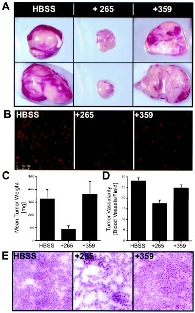
Figure 5.
TSRI265 suppresses tumor growth on the chick CAM via impairment of angiogenesis. (A) Primary tumors were grown on the CAMs of 9-day embryos by implantation of 5 × 10⁶ CS-1 cells and incubation for 7 days. At this point, 50-mg sections of these tumors were subcultured onto fresh 9-day CAMs. After 24 h, embryos were injected i.v. with 100 μl of 100 μM (≈10 μg) TSRI265 or TSRI359, or Hanks' balanced salt solution vehicle alone. Tumors were harvested 10 days later, trimmed of adjacent stromal tissue, and weighed wet (C). (B and D) Quantification of blood vessel density in treated and control tumors. (B) Tumors harvested as above were snap frozen, sectioned, and stained with anti-vWF polyclonal antibodies and an Alexa 568-labeled secondary antibody. Representative anti-vWF staining of treated and control tumors. (Bar = 100 μm.) (D) Quantification of the number of blood vessels per field as defined by anti-vWF reactivity. Data shown are the mean ± SE. (E) Serial sections of tumors were stained with hematoxylin and eosin for standard histological analysis. Representative photomicrographs are shown.

