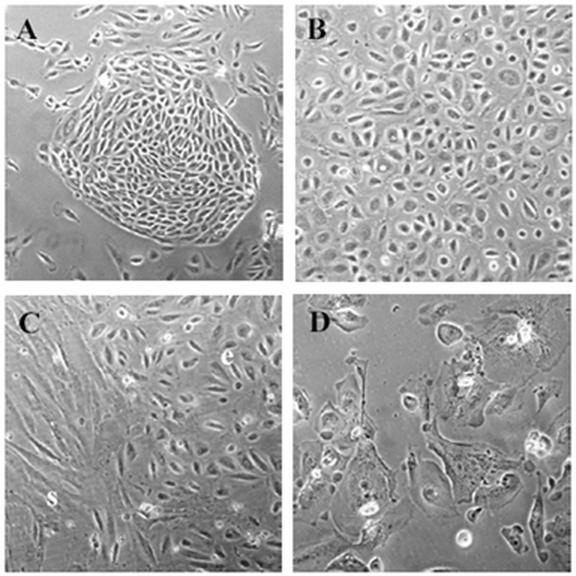
Fig. 1.

Primary human ovarian surface epithelium (OSE) morphology. A) Nest of OSE cells 4 days after initial plating. B) Monolayer of ‘cobblestone’ OSE at passage 1. C) Stromal cell (s) contamination of OSE (e) culture. D) Senescent OSE cells at passage 4. A = 100X magnification, B-D = 200X.
