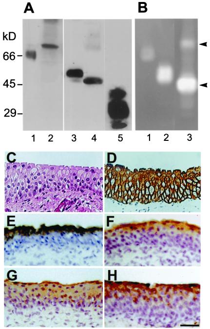
Figure 2.
Detection of PAs and PP5 in bovine urothelium. (A) Immunoblot analysis. Total bovine urothelial proteins (10 μg; lanes 2, 4, and 5), recombinant human (rh) tPA (66 kDa, 0.2 μg; lane 1), or rh-uPA (52 kDa, 0.2 μg; lane 3) produced in bacteria were separated by SDS/PAGE and immunoblotted with antibodies against tPA (1 and 2), uPA (3 and 4), and PP5 (5). Note the detection of bovine tPA (68 kDa), uPA (48 kDa), and PP5 (27, 31, and 35 kDa) in bovine urothelial extracts; the three PP5 bands reflect different degrees of glycosylation (ref. 50 and data not shown). The sizes of the human and bovine PAs are known to be slightly different. (B) Detection of fibrinolytic activities of rh-tPA (lane 1, 0.1 μg), rh-uPA (land 2, 0.1 μg), and a total bovine urothelial extract (lane 3, 10 μg) by zymography. The SDS gel was overlaid with a fibrin-containing indicator gel, incubated at 37°C for 1–3 h, followed by Coomassie blue staining of the indicator gel. Note the bovine tPA (68-kDa) and uPA (48-kDa) bands (arrowheads). (C–H) Immunolocalization of the PAs and PP5. Paraffin sections (5 μm) of bovine bladder were stained with hematoxylin and eosin (C), AE1 and AE3 antibodies to keratins (D) (16, 51), rabbit antitotal uroplakins (E) (4), mouse anti-tPA (F), rabbit anti-uPA (G), and rabbit anti-PP5 (H) (50). Note the staining of superficial cells by anti-tPA and anti-PP5 and the suprabasal cells by anti-uPA. (C–H) Same magnification. (Bar = 50 μm.)

