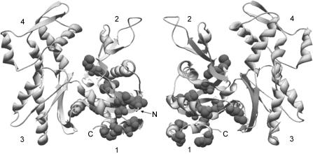
Figure 10. Muscle specific substitutions in subdomain 1 of yeast actin.
Muscle specific substitutions in subdomain 1 of yeast actin are depicted as space-filled residues in the ribbon structure of yeast actin (protein data base identification: 1YAG, Vorobiev et al. 2003). Both faces of the actin molecule are shown and the N- and C-termini are marked where visible. The numbers denote the four actin subdomains. ATP housed in the cleft has been omitted from the structure.

