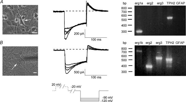
Figure 3. Transcripts for different erg channels are present in embryonic rhombencephalon neurones as revealed by single-cell RT-PCR.
Brief electrophysiological recordings in 40 mm K+ bath solution were performed prior to single-cell RT-PCR. Voltage pulse protocol shown at the bottom. Holding potential was −20 mV. Erg currents were activated by a voltage pulse to +20 mV for 2 s and inward currents were elicited by 100 ms pulses to potentials from −90 to −120 mV in steps of 10 mV. Erg currents were identified by the typical hook-like shape. A, large, multipolar neurone (left, arrow) from which transient inward currents typical for the presence of erg current were recorded (centre). Single-cell RT-PCR for this cell (right) in which erg1a and TPH2 were found. B, large, multipolar neurone (left, arrow) from which an erg current was recorded (centre). Transcripts for all three tested erg channel subunits were found in this cell (right). The absence of glial fibrillary acidic protein (GFAP), normally present in astrocytes, served as a negative control. Scale bars: 10 μm.

