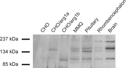
Figure 5. Different erg1 splice variants are expressed in rhombencephalon as revealed by Western blot.
Immunoblots of membrane preparations of untransfected CHO cells, CHO cells transiently expressing erg1a or erg1b channels, lactotrope MMQ cells, rat pituitary, rat embryonic rhombencephalon and adult rat brain. An antibody against the erg1 C-terminus was used. There was no staining in untransfected CHO cells. The antibody detected proteins in CHO cells expressing erg1a or erg1b at positions expected for erg1a and erg1b. Proteins close to the size of erg1a in CHO cells were found in MMQ cells, pituitary, rhombencephalon and brain. The bands with slightly higher molecular mass probably represent the glycosylated proteins. Proteins close to the size of erg1b in CHO cells were found in MMQ cells, pituitary, rhombencephalon and brain, representing glycosylated forms of erg1b. In addition, in MMQ cells, pituitary, rhombencephalon and brain the antibody stained a high molecular mass protein (∼200 kDa) of unknown origin.

