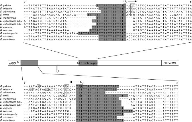
Figure 6.
The nucleotide sequences around the T-stretches and the positions of the OR of Drosophila mtDNA. The nucleotide sequences for several Drosophila species that were determined thus far were aligned with the T-stretches (shaded areas) on the major coding (top) and the minor coding (bottom) strands, respectively. The sites where the free 5′ ends were mapped for four Drosophila species in this study are indicated by boxes. The direction of replication is indicated by an arrow. The stem-loop structure previously proposed is also shown (Clary and Wolstenholme 1987; Monforte et al. 1993).

