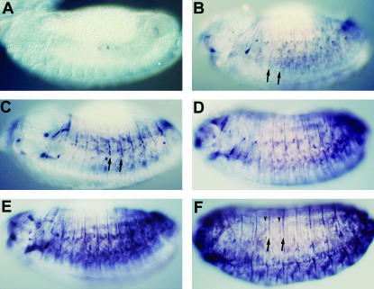
Figure 2.
Developmental profile of stv embryonic expression revealed by whole-mount anti-STV staining. All views are lateral. (A) A stage 13 embryo showing the earliest detectable STV expression, after germ-band retraction has been completed. (B) A mid-stage 13 embryo. STV localization is much more extensive, although very faint compared with older embryos. Intersegmental stripes (arrows) can be seen, in addition to spots of localization. (C) A late stage 13 embryo. Staining is darker, especially in the intersegmental stripes (arrows), and the level of intrasegmental staining is increasing. (D and E) Early and late stage 14 embryos. (F) The mature embryonic STV expression pattern at stage 15. This corresponds to the stage at which most muscles have formed their attachments with the epidermis. STV is present in intersegmental (arrows) and intrasegmental (arrowheads) attachment cells and in muscles.

