Figure 3.
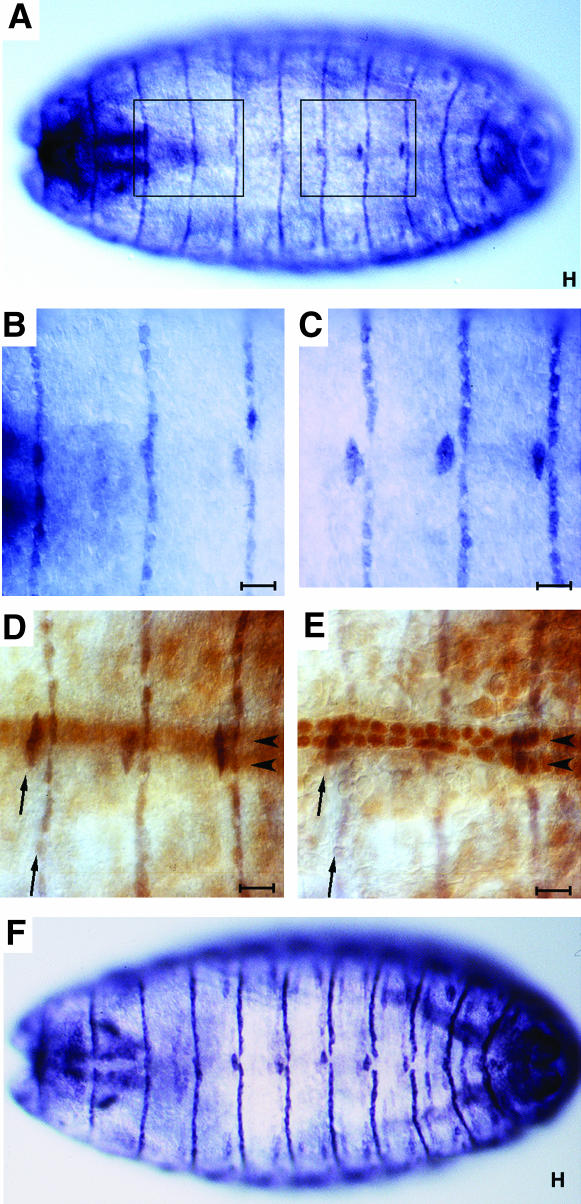
STV is present in all dorsal epidermal tendon cells. All panels are dorsal views of stage 16 embryos. (A) Anti-STV staining reveals one stripe of STV per segment, extending across the dorsal epidermis of the embryo. Five of these stripes, those at the anterior of segments A2–A6, are broken at the dorsal midline. (B) Higher magnification of the anterior segments boxed in A showing stripes anterior to segments T3, A1, and A2. Only that of A2 is broken at the dorsal midline. (C) Higher magnification of the posterior segments boxed in A showing three stripes of STV localization which are all broken at the dorsal midline (anterior of segments A4–A6). (D) A higher-magnification view of a howE7-3-4 lacZ embryo, an enhancer trap that expresses strongly in nuclei of dorsal vessel cells that flank the dorsal midline. The darker staining in focus shows STV expression (arrows) while the lighter brown color is anti-βGal staining of the dorsal vessel nuclei (arrowheads, out of focal plane). (E) The same embryo with the dorsal vessel nuclei staining in focus and STV staining out of focus. The STV stripes have a width of one cell and the broken portion of the stripe represents two cells, one on either side of the dorsal midline, which are displaced one cell from the main stripe. (F) Anti-Alien staining, showing the same pattern as STV localization. Alien is present in epidermal tendon cells (Goubeaud et al. 1996). All bars, 10 μm.

