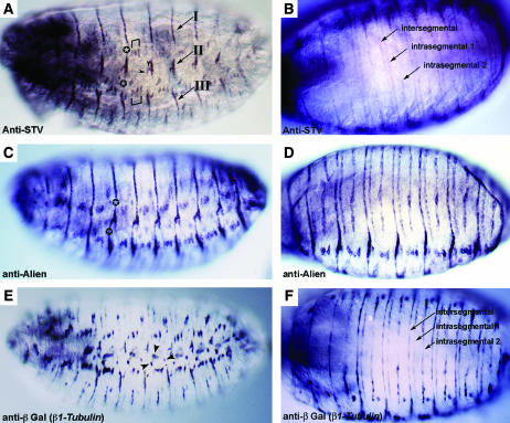
Figure 4.
STV is present in lateral epidermal and ventral epidermal tendon cells. All embryos are at stage 16. (A, C, and E) Lateral views. (B, D, and F) Ventral views. (A and B) Anti-STV staining. (A) The stripes of STV localization that are observed dorsally (Figure 3) are disrupted laterally. The dorsal stripes form attachment group I. STV is also present in the short lateral stripe representing group II and the more ventral group III. Brackets mark the dorsal and ventral attachments of muscles LT1 (21), LT2 (22), and LT3 (23). Arrowheads indicate the ventral attachment of muscle DT1 (18) and the posterior attachment of muscle DO5 (20). Other single attachment points cannot be readily identified. (B) Three stripes of STV localization per segment are observed ventrally. Morphologically, these stripes will generate three furrows, or apodemes: one intersegmental and two intrasegmental (Campos-Ortega and Hartenstein 1997). (C and D) Anti-Alien staining. (C) Alien expression differs from STV expression by continuing across the lateral regions among the attachment groups I, II, and III (indicated by a star). (D) A ventral view shows a pattern identical to that of STV. (E and F) Anti-βGal staining of an embryo carrying a β1Tubulin∷lacZ construct. (E) lacZ is expressed in all tendon cells plus the chordotonal organs (arrowheads). Like STV, β1Tubulin expression is absent among the attachment groups. (F) β1Tubulin is expressed in the same three stripes per segment as STV and Alien.

