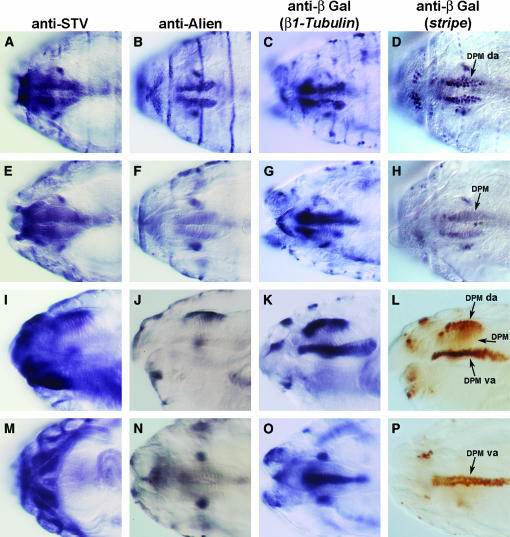
Figure 5.
The pattern of STV expression in the stage 16 embryonic head. A, E, I, and M are stained with anti-STV. B, F, J, and N are stained with anti-Alien. (C, G, K, and O) Embryos carrying a β1Tubulin∷lacZ promoter construct stained with anti-β̃galactosidase. (D, H, L, and P) Embryos carrying an enhancer trap inserted in stripe and stained with anti-β-galactosidase. (A–H) Dorsal views. (A) STV is present in the dorsal attachment of the DPM. STV is expressed in essentially the same pattern as Alien, β1Tubulin, and Stripe markers (B–D), confirming that STV is expressed in tendon cells. (E–H) The same embryos shown in A–E viewed in a different focal plane, focused on the DPM, showing staining for STV to be out of focus and confirming that STV is present in the attachment cells of the DPM. (I–L) Lateral views. (I) STV is localized to the dorsal attachments of the DPM, but not to the DPM itself. The ventral attachment cells of the DPM did not stain. (J–L) Alien, β1Tubulin, and Stripe expression, respectively. Dorsal attachment but not ventral attachment localization is evident for Alien, while β1Tubulin and Stripe are present in both dorsal and ventral attachments of the DPM. (M–P) Ventral views. STV is expressed strongly in the large ventral intersegmental muscles (VIS), but Alien (N), β1Tubulin (O), and Stripe (P) are not. The stripe enhancer trap clearly shows the two rows of cells that make up the ventral attachments of the DPM. DPM da, dorsal attachment of DPM; DPM va, ventral attachment of DPM.

