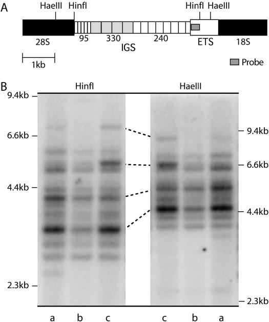
Figure 3.
IGS length profiles reproduced with two different restriction enzymes. (A) A diagram of the IGS region showing restriction sites and probe location. The IGS is mostly composed of tandem repeats of 95, 330, and 240 bp. (B) Genomic DNA was digested with HinfI or HaeIII, fractioned through a 1% agarose gel, and transferred to nitrocellulose. The resulting Southern blot was probed with a region of the ETS, marked with a shaded bar in A. The position of DNA standards in kilobases is indicated on the left and right. Lanes labeled a contain DNA from Harwich line 3, lanes labeled b contain DNA from line 20, and lanes labeled c contain DNA from line 2. Several corresponding bands in these last two lanes are connected with lines. The pattern of IGS bands is identical between the HinfI digests and HaeIII digests, with HinfI producing smaller fragments that are better separated on the gel.

