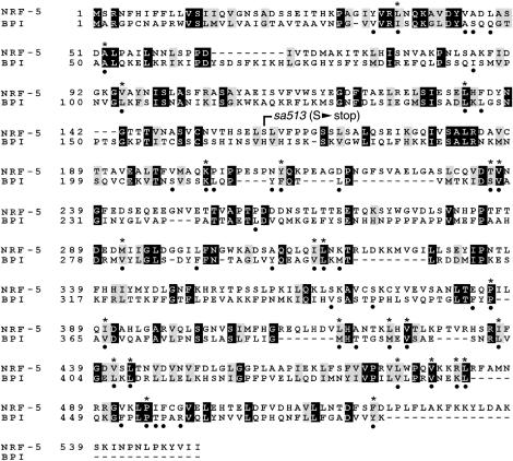
Figure 3.
Alignment of nrf-5 and human BPI protein. Solid boxes denote identical amino acids; shaded boxes denote similarities. The position of the mutation S159 to stop in sa513 is indicated. Solid dots below the alignment indicate 47 phospholipid contact residues based on BPI crystal structure (Beamer et al. 1997). Asterisks above the alignment indicate the 25 identical or similar residues in nrf-5 among these 47 contact residues.

