Figure 3.
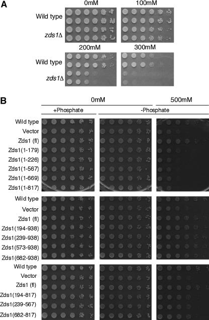
Calcium sensitivity of zds1-disrupted cells. (A) SP66 (h90, wild type) and MY6010 (h90, zds1Δ) cells were cultured at 30° in liquid medium until they reached log phase. They were concentrated to 2 × 10⁷ cells/ml and then diluted sequentially fivefold (to the right direction). The cells were spotted on YEA plates containing 0–300 mm CaCl2 and incubated at 30° for 3 days (0 and 100 mm CaCl2), 4 days (200 mm CaCl2), or 7 days (300 mm CaCl2). (B) The C-terminal region of Zds1 suppresses the calcium sensitivity of zds1-disrupted cells. SP66/pSLF272L GFPS65A cells were spotted on the plate as a wild-type strain. All other strains were transformants of MY6010. All plasmids were derived from the vector pSLF272L GFPS65A. The transformants were cultured at 30° in liquid PMA medium containing thiamine until they reached log phase, after which they were washed to remove the thiamine and resuspended in H2O. The cell concentration was adjusted to 2 × 10⁷ cells/ml and the cells were diluted sequentially fivefold (to the right direction). Cells were spotted on PMA (+phosphate), PMA containing sodium acetate instead of sodium phosphate (−phosphate; middle), or PMA (−phosphate; right) containing 500 mm CaCl2. These plates were incubated at 30° for 5 days (without CaCl2) or for 8 days (with CaCl2).

