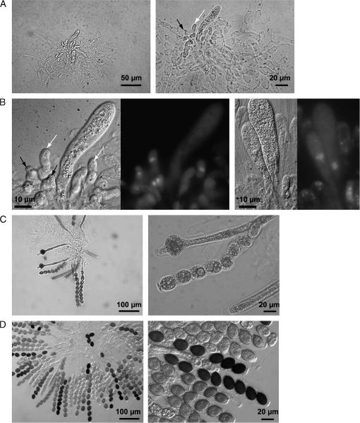
Figure 5.
Microscopic analysis of ascus development in the rarely produced perithecia of the Δpre2/Δppg2 double-mutant strain. (A) Sixty percent of all squeezed perithecia contain few hook cells (solid arrow) and undifferentiated asci (ascus initial, open arrow). (B) DAPI staining identified nuclei during crozier (solid arrows) and ascus formation (ascus initial, open arrow). (C) Thirty percent of ascus rosettes carry 3–20 more or less differentiated asci with ascospores. (D) Ten percent of perithecia contents represent normally developed asci with ascospores.

