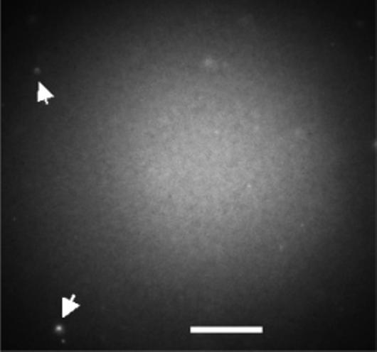
Figure 1.

Fluorescence image of a POPC/NBD-PC bilayer formed on an ATRP polyacrylamide-modified fused silica surface. The arrows point to unfused lipid vesicles either in solution (diffusing rapidly) or attached to the surface (diffusing slowly). Excitation is with a mercury lamp. The scale bar is 70 μm.
