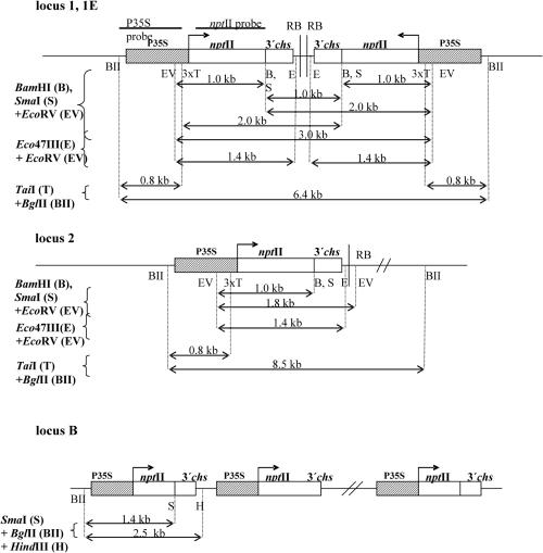
Figure 1.
Schematic representation of the genomic organization of transgenic loci and physical maps of the restriction sites. The transgenic locus 1 has been previously reported as locus X (29). The methylation analysis of the promoter involved the three TaiI sites; the diagnostic sites for the analysis of the nptII-transcribed region were SmaI and BamHI, and Eco47III for the non-transcribed sequences at the right border. Evidence for the IR character of the T-DNA insertions in locus 1 and locus 1E has been given elsewhere (18). EcoRV, BglII, and HindIII enzymes were used to dissect particular subregions of T-DNA. P35S, promoter of the cauliflower mosaic virus; nptII, neomycin phosphotransferase II gene; RB, T-DNA right border; 3′chs, transcription termination sequence from the 3′-untranslated region of the chalcone synthase gene from snapdragon (Antirrhinum majus).

