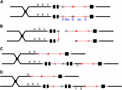
Figure 1.
Model for formation of deletions by sister-chromatid transposition. (For animated version, see supplemental material at http://www.genetics.org/supplemental/.) The diagram pertains to the structure of the p1-vv9D9A allele, which is the progenitor of the p1-ww deletion alleles described in the text. The two lines indicate sister chromatids joined at the centromere, which is indicated by a solid circle. The solid black boxes indicate the three exons of the p1 gene; the 5′-end of the p1 gene is closer to the centromere (Zhang and Peterson 1999). The red arrows indicate the Ac or fAc elements inserted into the second intron of the p1 gene, and the open and solid arrowheads indicate the 3′- and 5′-ends, respectively, of Ac/fAc. The short black line between Ac and fAc indicates a 112-bp rearranged p1 sequence (rP) present in the p1-vv9D9A allele (not to scale). (A) Following DNA replication, identical sister chromatids are joined at the centromere. Ac transposase (small circles) binds to the 5′ terminus of Ac in one chromatid and to the 3′ terminus of fAc in the sister chromatid. (B) Cuts are made at the Ac and fAc termini to excise the transposon ends. The two nontransposon ends join together at the site marked by the black X to form a chromatid bridge. (C) Reinsertion of the excised transposon ends into the chromatid bridge between b and c generates two reciprocal chromatids; one carries a deletion of c and the other carries an inverted duplication of c. (D) Same as for C, except that reinsertion between a and b generates one chromatid with a deletion of b c and one with an inverted duplication of b c. For simplicity, the model depicts fully replicated sister chromatids at the time of transposition. In reality, transposition may occur when the chromosomes are partially replicated.

