
Figure 3.
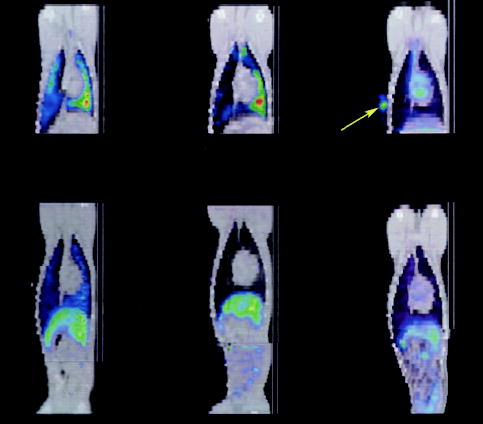
Coronal fused SPECT (color) and CT (gray-scale) image of a dog with (top left) and without (top middle) MI during the first hours after intravenous injection of 111In oxine–labeled MSCs showing predominant lung uptake with increased uptake to the dependent left lung (green-yellow color toward right). In a dog with MI that received 111In oxine without MSCs (top right), the tracer behaves primarily as a blood pool agent, with uptake visible in left and right ventricles of the heart. A reference marker (arrow) containing 111In oxine was placed on the chest wall on the dog that did not receive MSCs (top right). Redistribution of 111In oxine–labeled MSCs to predominantly the liver occurs at 24 hours after intravenous injection in both a representative infarcted (bottom left) and noninfarcted (bottom middle) dog. In an infarcted dog injected intravenously with 111In oxine only (ie, no MSCs), a similar pattern of redistribution to the liver is observed at 24 hours after injection (bottom right).