
Figure 8.
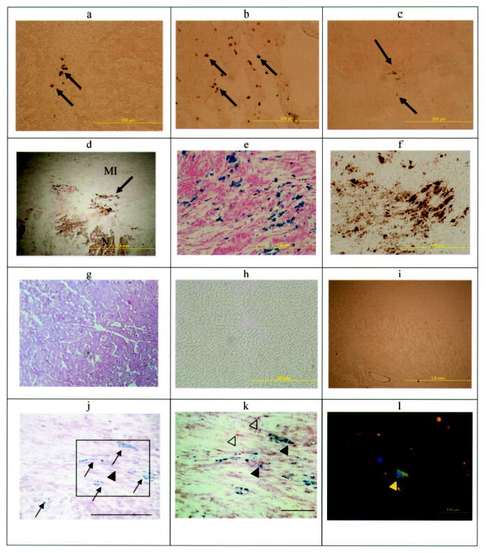
Photomicrographs of various organs demonstrating the presence or absence of Feridex-labeled MSCs. DAB-enhanced Prussian blue–stained photomicrographs of liver (a), lung (b), spleen (c), and heart (d) demonstrate iron-positive cells in these organs. Higher magnification of d showing Prussian blue staining with (e) and without (f) DAB enhancement demonstrates the intracytoplasmic iron and MSC localization in the MI rim (N indicates normal myocardium). No iron-positive cells were demonstrated with or without DAB-enhanced Prussian blue staining in the noninfarcted myocardium (g, h) or kidney (i). j, Double-staining cells for acid phosphatase (red) and iron (blue) indicate that macrophages (arrowhead) in the heart were rare, whereas the majority of Prussian blue–positive cells were the original Feridex-PLL–labeled MSCs (arrows) (bar = 200 μm). k, Inset of j at a higher magnification demonstrates primarily MSCs (blue stain) with a few macrophages with (filled arrowheads) and without (open arrowheads) iron (bar = 100 μm). l, Dextran staining (green, yellow arrow) indicates Feridex retention in a labeled MSC in the heart. Nuclei are stained with DAPI (blue).