Figure 3.
Tissue Expression and Subcellular Localization of THF1.
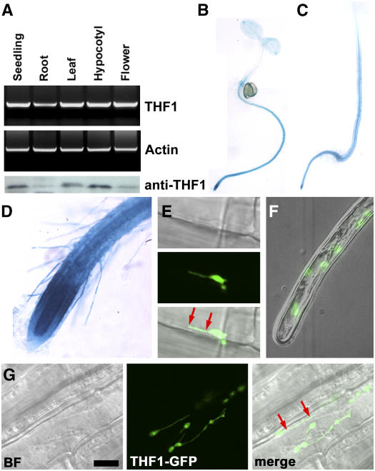
(A) THF1 transcript and THF1 protein in different organs. Top, RT-PCR using RNA isolated from the indicated organs from 30-d-old light-grown plants and 10-d-old seedlings. Bottom, the same set of samples was used for immunoblot analysis probed with anti-THF1 serum (α-THF1). The lowest and highest signals were in the linear range of this assay.
(B) and (C) Transcriptional fusions between the THF1 promoter and the uidA gene encoding GUS were used to transform Arabidopsis. Seedlings were stained for GUS activity in 5-d-old seedlings grown either in constant light (B) or in darkness (C). Staining was stopped after 5 h to highlight the predominant expression locations, but overnight staining indicated ubiquitous expression.
(D) Root tip cells including hairs express THF1; the strongest expression was seen in the root meristem.
(E) to (G) A 35S-driven THF1-GFP fusion protein localizes to plastids and can be seen in plastid stromules that appear to tether the plastid to the plasma membrane (red arrows; see also Supplemental Movie 1 online). (E) and (G) show root epidermal cells; (F) shows a root hair cell. BF, bright field. Bar in (G) = 10 μm.

